|
|
|
| Evaluation Of The Use Of Gothic Arch Tracing Width As A Guide For Positioning Of Artificial Teeth: A Clinical Study |
Rohit U Nair 1 , Dhanyakumar B.H. 2 , Nandeeshwar D.B. 3 , Chetan Pathak 4
1 Lecturer, Dept. of Prosthodontics - YMT Dental College & Hospital,Navi Mumbai
2 Professor, Dept. of Prosthodontics - Bapuji Dental College & Hospital, Davangree, Karnataka, India
3 Professor and HOD, Dept. of Prosthodontics - Bapuji Dental College and Hospital, Davangere, Karnataka, India
4 Senior Lecturer, Department of Prosthodontics - Sudha Rustagi College of Dental Sciences & Research, Faridabad, Haryana, India
|
| Address For Correspondence |
Dr. Rohit U Nair
Lecturer, 13, Department of Prosthodontics
YMT Dental College & Hospital Kharghar,
Navi Mumbai - 410210 Maharashtra, India.
Email ID - rohit302@gmail.com
Phone no.: +91 9004242413 |
| Abstract |
| Purpose : Several guidelines for positioning of artificial teeth in complete denture fabrication exist. Anatomical landmarks, although much discussed, are not solely reliable indicators of tooth position. Scant information is present in the literature regarding the use of mathematical ratios for arrangement of artificial teeth. The purpose of this investigation was to determine if any relationship existed between the width of a Gothic arch tracing and the intercanine width in an Indian population.
Materials and Methods : Twenty subjects participated in this study and performed mandibular movements to record an arrow-point tracing on an intraoral tracing plate using a stylus embedded in a palatal plate made of autopolymerizing acrylic resin. The width of the tracing was measured. The intercanine width of the subject determined from an accurate diagnostic cast was divided by the tracing width to obtain a ratio.
Results : A mean ratio of 2.13 (s.d. = 0.26) was calculated for the given sample. No significant differences were seen between the male and female groups (p<0.05).
Conclusions : The width of the Gothic arch tracing can probably be used as in indicator for artificial tooth positioning.Results of this study may be tested in a larger study sample to evaluate reliability for use as a mathematical index for anterior teeth arrangement. |
|
| Keywords |
| Ratio, Mathematical Index, Intercanine Width, Gothic Arch Tracing Width, Intraoral Tracing, Arrow-point tracing |
|
| Full Text |
Introduction
The arrangement of artificial teeth during complete denture fabrication constitutes a critical decision-making step in prosthodontics in that, objective scientific principles must be followed to realize the patient’s largely subjective expectations of what the eventual appearance of the prosthesis might be. Pre-extraction records such as good quality photographs and diagnostic casts, all of which provide valuable information about previous tooth positions, are rarely available to the attending operator, except for when the patient has previously received an immediate denture prosthesis. As a result, the task of first selecting and then correctly arranging the artificial teeth is made difficult.
While several methods are available to aid in selecting the “correct” tooth mold[1],[2],[3],[4],[5], artificial teeth arrangement remains, for the most part, guided by either anatomical landmarks in the immediate vicinity of the denture teeth[6], or by means of prefabricated templates[7],[8], or even by arbitrary means based on one’s experience and idea of a final esthetic result.[9]
Amongst the various anatomical landmarks, the incisive papilla has been the subject of much study with regards to its validity as a stable landmark for artificial teeth arrangement.[10],[11],[12],[13] However, the shape, positional relation to natural teeth prior to extraction and post-extraction position of the papilla has been shown to be variable. Latta and others, while investigating the reliability of some routinely used facial measurements in 109 edentulous subjects, concluded that the inter-alar width, inter-pupillary distance and bi-zygomatic width show great variations and, consequently, are unreliable for use as guides for positioning denture teeth.[3]
El Gheriani and Winstanley correlated the distance between the side-arms of Gothic arch tracings obtained from 25 subjects of different races, with maxillary inter-cuspid distances measured from the same subject. Their findings showed that the inter-cuspid distance was almost twice the side-arm width of the tracing record.[14]
Further attempts at deriving mathematical relationships for tooth positioning have been made. The same authors examined the relationship between the distance between the buccal cusps of natural maxillary teeth and the side arms of a Gothic arch tracing and found a constant ratio between the measured variables.[15] When applied clinically, the authors found that 38 out of 40 patients who received dentures fabricated by using the mathematical index for teeth arrangement were more satisfied at the end of a 1-week trial, when compared with dentures in which teeth were arranged conventionally.[16]
Keshvad and Winstanley studied the relationship between the intercondylar width and interdental widths of the maxillary and mandibular canines and first molars. A strong correlation was found between the measured variables and a set of indices was developed from the results to be used for the positioning of complete denture teeth.[17]
The purpose of the present study was to determine if there was any relationship between the width of the side-arms of the Gothic arch tracing (GATW) and the inter-arch distance between the maxillary canines in an Indian population (ICW).
Materials and Methods
20 adult Indian subjects (10 males & 10 females) aged 19-29 years were selected for this study. All subjects had a full complement of natural teeth (up to 2nd molars), with no history of orthodontic treatment, orthognathic surgery, Temporomandibular Joint (TMJ) pain or dysfunction, no full veneer restorations on the maxillary canine teeth and no evidence of attrition or periodontal disease.
Maxillary and mandibular full-arch impressions were made using irreversible hydrocolloid impression material (Zelgan, Dentsply India) in perforated rim-lock stock metal impression trays (Addler Co., Germany). The impressions so obtained were then poured in dental stone (White Gold Dental Stone; Rajkot, India) to obtain diagnostic casts.
A palatal plate was fabricated using autopolymerizing acrylic resin (RR; DPI, Mumbai, India) by the sprinkle-on technique on the maxillary cast and a stylus with an adjustable height-screw (Fig. 1-A) was embedded on the plate using heated modeling plastic impression compound (Pinnacle; DPI, Mumbai, India) such that the point of the stylus rested on the intersection of an imaginary line passing between the first and second premolars on each side and the midline of the cast (Fig. 1-B). A metal intraoral tracing plate was then coated with soot to serve as the tracing medium. The plate was immobilized intraorally by means of heated modeling plastic impression compound placed laterally on the under-side so that when pressed against the occlusal surface of the mandibular teeth and cooled, it would maintain the stability of the plate while the tracing procedure was in progress (Figs. 1-C, D).
Intraorally, the stylus screw was adjusted to eliminate occlusal contacts between opposing natural teeth during lateral and protrusive movements with minimal tooth separation. The subjects were then instructed to move the mandible from a retruded position forward into protrusion and then back again. Lateral movements of the mandible were then introduced and the subjects were asked to make excursions to both right and left extremes of mandibular movement (Figs. 2-A, B, C).
 | Figure 1: Preparation Of Gothic Arch Tracing Plates: 1a. Stylus / Central Bearing Point And Intraoral Tracing Plat, 1b. Stylus Attached To Palatal Plate Made Of Autopolymerizing Acrylic Resin Using Modeling Plastic Impression Compound, 1c. Intraoral Traci
 |
 | Figure 2: Mandibular Movements Performed By Subject: 2a. Maximum Right-lateral Excursion, 2b. Maximum Protrusion, 2c. Maximum Left-lateral Excursion
|
The plate bearing the arrow-point tracing was then retrieved from the mouth and a digital caliper (Zoom, accurate up to 0.01mm) was used to make the following measurements (in millimeters):
1. The distance between the ends of the right and left arms of the Gothic arch tracing (Fig. 3-A).
2. On the maxillary cast, the distance between the lingual inclines of the tips of the natural canine teeth (Fig. 3-B).
Mean values for ratio between intercanine width and Gothic arch tracing width were then calculated.
 | Figure 3: Measurements : 3a. Distance Between End-points Of Lateral Arms Of Tracing Measured In Millimeters Using A Digital Caliper, 3b. Distance Between Lingual Inclines Of Cusp Tips Of Maxillary Canine Teeth On Diagnostic Cast Measured In Millimeters Us
|
Results
The results are presented in Table 1. The intercanine widths of the subjects were divided by the distance between the arms of the Gothic arch tracing and in each case a ratio was obtained. Male subjects showed a mean ratio of 2.18 (S.D. = 0.30) as against females who recorded a mean ratio of 2.07 (S.D. = 0.23). The mean ratio for both sexes was 2.13 (S.D. = 0.26). No significant differences were found between the mean ratios obtained for male and female subjects (Mann Whitney U = 36, n1 = n2 = 10, P < 0.05 two-tailed) (Fig. 4). To examine the correlation between the maxillary intercanine widths and the Gothic arch tracing widths, a Spearman’s rank correlation coefficient test was performed (Fig. 5). A moderate positive correlation was found to exist between the measured variables for the given sample (r=0.23).
 | Figure 4: Ratio Of Maxillary Inter-canine Width To Gothic Arch Tracing Width
|
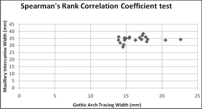 | Figure 5: Spearman's Rank Correlation Coefficient Test
|
Discussion
The use of tracing devices to record a Gothic arch was first suggested by Gysi in the early 1900s.[18] The Gothic arch tracing represents the border movements of the mandible in the horizontal plane with its apex indicating the most retruded position of the mandible.[19] Its principal application in prosthodontics has been to verify the centric relation position recorded at the time of the jaw relation appointment. While this practice continues to hold on, few attempts have been made to obtain additional information from the tracing itself. This study has attempted to evaluate any relationship that might exist between the intercanine width of the maxillary arch and the width of the subject’s Gothic arch tracing.
As seen from (Table 1). a mean ratio of 2.13 was found to exist between the previously mentioned variables. No significant differences were found to exist between the mean values for male and female subjects. This result is in agreement with those of El Gheriani and Winstanley where it was found that the Gothic arch tracing width was on an average half of the maxillary intercanine width (mean ratio : 2.04).[14] In that study, Caucasian, Malaysian and Arabic subjects were assessed using a similar methodology and mean ratios for ICW:GATW of 1.9, 1.98 and 2.2 were obtained respectively.[14] The authors did not group subjects based on sex and consequently any differences that might have existed therein were not statistically analyzed.
 | Table 1
|
In this study, a positive correlation, although weak (r=0.23), was found to exist between the measured variables suggesting that the Gothic arch tracing width increased with increases in the intercanine distance, contributing to the similar ratios obtained for a majority of the subjects. Further studies are required to validate and generalize the results of this study by considering a larger and more diverse sample of the Indian population. Additionally, other variables such as effect of arch form on the derived ratio may also be investigated.
Although not comparable, it may be noted that mathematical indices or ratios of the type obtained here have been derived independently, and tested for reliability, by Keshvad and Winstanley while studying the relationship between the intercondylar and intercanine widths.[17]
Conclusion
Within the limitations of this study, it can be concluded that a mean ratio of 2.l3 exists between the maxillary intercanine width and the width of a Gothic arch tracing obtained from subjects of an Indian population.
References
1. Scandrett FR, Kerber PE, Umrigar ZR. A clinical evaluation of techniques to determine combined width of the maxillary anterior teeth and the maxillary central incisor. J Prosthet Dent 1982;48: 15-22.
2. Hoffman W, Bomberg TJ, Hatch RA. Interalar width as a guide in denture tooth selection. J Prosthet Dent 1986;55: 219-221.
3. Latta GH Jr., Weaver JR, Conkin JE. The relationship between the width of the mouth, interalar width, bizygomatic width, and interpupillary distance in edentulous patients. J Prosthet Dent 1991;65: 250-254
4. Varjao FM, Nogueira SS. Intercommissural Width in 4 Racial Groups as a Guide for the Selection of Maxillary Anterior Teeth in Complete Dentures. Int J Prosthodont 2005;18: 513-515.
5. Varjao FM, Nogueira SS. Nasal Width as a Guide for the Selection of Maxillary Complete Denture Anterior Teeth in Four Racial Groups. J Prosthodont 2006;15: 353-358.
6. Roraff AR: Arranging artificial teeth according to anatomical landmarks. J Prosthet Dent 1977;38: 120-130
7. Beresin VE, Schiesse FJ: The neutral zone in complete dentures. J Prosthet Dent 1976;36: 356-367
8. Stananought D: The setting of teeth, in Newton AV (ed): Laboratory Procedures for Full and Partial Dentures, ed 1. London, Blackwell Scientific Publications, 1978, pp 139-140
9. Waugh DB: The arrangement of teeth in the natural and artificial dentures. Dental Cosmos 1936;78: 1125-1135
10. Harper RN: The incisive papilla – The basis of a technic to reproduce the positions of key teeth in prosthodontia. J Dent Res 1948;27: 661-668
11. Mersel A, Ehrlich J: Connection between incisive papilla, central incisor and rugae canine. Quintessence Int 1981; 12: 1327-1329
12. Grave AM, Becker PJ: Evaluation of the incisive papilla as a guide to anterior tooth position. J Prosthet Dent 1987; 57: 712-714
13. Grove HF, Christensen LV: Relationship of the maxillary canines to the incisive papilla. J Prosthet Dent 1989;61: 51-53
14. El-Gheriani AS, Winstanley RB. The value of the Gothic arch tracing in the positioning of denture teeth. J Oral Rehab 1988;15:367-371
15. El-Gheriani, Davies AL, Winstanley RB. The Gothic arch tracing and the upper canine teeth as guides in the positioning of upper posterior teeth. J Oral Rehab 1989;16:481-490
16. El-Gheriani AS. A new guide for positioning of maxillary posterior denture teeth. J Oral Rehab 1992; 19: 535-538
17. Keshvad A, Winstanley RB, Hooshmand T. Intercondylar width as a guide to setting up complete denture teeth. J Oral Rehab 2000;27:217-226
18. Gysi A. The problem of articulation. Dent Cosmos 1910; 52: 1-19, 148-169, 268-283, 403-404
19. Johnson T. Gothic arch tracing devices. Quintessence Dent Technol 2006;4: 130-143.
|
|
|
|
|
|
|